Ya-Feng Liu ![]() ,
Rui Liu,
Xue-Yang Li,
Zhen Song,
Xue-Hang Zhao
,
Rui Liu,
Xue-Yang Li,
Zhen Song,
Xue-Hang Zhao
For correspondence:- Ya-Feng Liu Email: liuyafeng68@hotmail.com Tel:+8637125550069
Received: 10 February 2016 Accepted: 18 June 2016 Published: 31 July 2016
Citation: Liu Y, Liu R, Li X, Song Z, Zhao X. Development of docetaxel and alendronate-loaded chitosan-conjugated polylactide-co-glycolide nanoparticles: In vitro characterization in osteosarcoma cells. Trop J Pharm Res 2016; 15(7):1353-1360 doi: 10.4314/tjpr.v15i7.1
© 2016 The authors.
This is an Open Access article that uses a funding model which does not charge readers or their institutions for access and distributed under the terms of the Creative Commons Attribution License (http://creativecommons.org/licenses/by/4.0) and the Budapest Open Access Initiative (http://www.budapestopenaccessinitiative.org/read), which permit unrestricted use, distribution, and reproduction in any medium, provided the original work is properly credited..
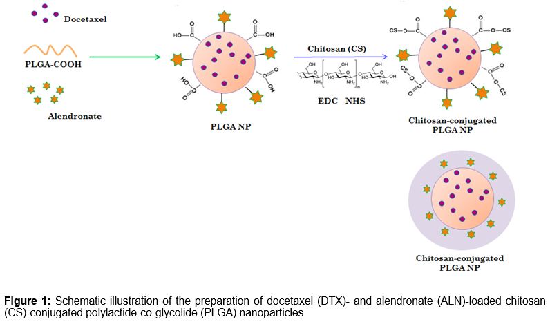
Purpose: To develop docetaxel (DTX)- and alendronate (ALN)-loaded, chitosan (CS)-conjugated poly-lactide-co-glycolide (PLGA) nanoparticles (NPs) to increase therapeutic efficacy in osteosarcoma cells.
Methods: Drug-loaded PLGA NPs were prepared by nanoprecipitation and chemically conjugated by the carboxylic group of PLGA to the amine-bearing CS polymer. The nanocarrier was characterized by dynamic light scattering, transmission electron microscopy, scanning electron microscopy, and differential scanning calorimetry as well as by in vitro drug release and cell culture studies.
Results: NP size was within the tumour targeting range (~200 nm) with an effective positive charge (20 mV), thus increasing cellular uptake efficiency. Morphological analysis revealed clear spherical particles with uniform dispersion. The NPs exhibited identical sustained release kinetics for both DTX and ALN. CS-conjugated PLGA with dual-drug-loaded (DTX and AL) NPs showed typical time-dependent cellular uptake and also displayed superior cytotoxicity in MG-63 cells compared with blank NPs, which were safe and biocompatible.
Conclusion: Combined loading of DTX and ALN in NPs increased the therapeutic efficacy of the formulation for osteosarcoma treatment, thus indicating the potential benefit of a combinatorial drug regimen using nanocarriers for effective treatment of osteosarcoma.
Introduction
Osteosarcoma is the most prevalent malignant bone cancer. It commonly occurs in children and adolescents: approximately 400 cases per year are diagnosed in the United States alone [1]. Osteosarcoma mostly arises in the metaphyseal ends of long bones, and is characterized by malignant metastatic tumours that differentiate as cartilage and bone [2]. The survival rate is nearly 20 % with surgery; however, it increases to 70 % when combined with anticancer drug therapy. Thus, surgery and chemotherapy play a vital role in the treatment of osteosarcoma. A recently multi-drug regimen reportedly improved osteosarcoma treatment [3].
Combination therapy has been considered a promising strategy for improving therapeutic efficiency and minimising side effects [4]. The combination of two or more chemotherapeutic drugs may act synergistically in cancer cell suppression [5,6]. In this study, docetaxel (DTX) and alendronate (ALN) were selected as a unique drug combination for the synergistic treatment of bone cancers.
DTX is a frontline chemotherapeutic agent that is active against a wide spectrum of cancers. DTX acts on the microtubules of the malignant cell to suppress its proliferation, killing cancer cells by inducing a G2/M phase arrest of the cell cycle. Despite excellent therapeutic potential, DTX treatment results in severe adverse effects, including neutropenia, anaemia, and hypersensitivity reactions [7]. ALN is used because of its strong osteoprotective properties and its affinity for bone cancer cells. It belongs to the class of amino bisphosphonates (NBP), and is in the fourth class of the Biopharmaceutics Classification System (BCS) because of its poor solubility and low bioavailability [8,9].
Nanotechnology-based formulation is a promising alternative in cancer treatment. Poly-lactide-co-glycolide (PLGA) exhibits excellent biocompatibility and biodegradability, and has been extensively used as a carrier for drug delivery [10]. This system extravasates solid tumours into the tumour site by passive targeting via an enhanced permeability and retention effect (EPR). However, the PLGA nanocarrier itself is not sufficient. Surface modification of PLGA with certain hydrophilic polymers enables these nanoparticles (NPs) to increase their selectivity for cellular binding and internalization [11]. For example, chitosan (CS), a biocompatible and nontoxic cationic polysaccharide, is commonly used for surface modifications.
The aim of this study was to develop a CS-coated PLGA nanocarrier system for the combined delivery of DTX and ALN to osteosarcoma cells.
Methods
Materials
PLGA-COOH with a lactide to glycolide ratio of 50:50 and an average molecular weight of 15 kDa was purchased from Ji’nan Daigang Biological Co. Ltd (Shandong, China). CS (deacetylation degree, >84 %) was purchased from Zhanjiang Xinmao Chemical & Glass Company (Zhanjiang, China). DTX, ALN, and 3-[4,5-dimethylthiazol-2-yl]-2,5-diphenyltetrazolium bromide (MTT) were purchased from Sigma Chemical Co., USA). All chemicals were analytical grade and used without further purification.
Preparation of CS-coated PLGA NPs containing DTX and ALN
DTX-loaded PLGA NPs were prepared by solvent diffusion (nanoprecipitation) as previously reported [12]. The organic phase (acetone) containing DTX (15 mg) and ALN (15 mg) and PLGA (200 mg) in a 1:1:1 ratio (v:v) was slowly injected (0.5 mL/min) into 20 mL of aqueous phase containing Poloxamer 188 (0.5 % w/v) as a stabilizer. The resulting NPs were recovered by centrifugation at 25,000 rpm for 30 min and purified by dialysis against water and lyophilized for 48 h.
The freeze-dried PLGA nanoparticles were dispersed in phosphate buffered saline (PBS) solution (pH 6.0) under bath sonication at 37 °C. N-hydroxysuccinimide (NHS) and N-(3-dimethylaminopropyl)-N′ ethyl carbodiimide hydrochloride (EDC·HCl) was added to activate the carboxyl group of PLGA. Different concentrations of CS were added into the above solution and the reaction was allowed to continue for 24 h. The excess EDC, NHS, and unreacted CS were eliminated by centrifugation. A schematic illustration of the preparation of dual-drug-loaded CS-coated PLGA NPs is shown in .
Particle size and zeta potential measurement
The hydrodynamic diameter of the NPs was measured using a dynamic light scattering (DLS) technique. A Zetasizer Nano ZS (Malvern Instruments, Worcestershire, UK) was used to measure the size at room temperature with an angle of detection of 176°. Zeta potential measurements were performed with a detection angle of 17° and calculated using the Smoluchowski model. The samples were suitably diluted (200 mg/mL) with double-distilled water such that the mean count rate was approximately 300 kcps.
Morphological characterization
Morphological studies of the NPs were conducted using transmission electron microscopy (TEM) and field emission scanning electron microscopy (FE-SEM). TEM (H-7600; Hitachi, Japan) was performed by staining a small drop of the NP suspension with a 2 % phosphotungstic acid (PTA) solution. The stained sample was deposited onto a carbon coated copper grid and dried for 30 min at room temperature.
The samples were examined under an acceleration voltage of 100 kV in conventional TEM-mode. FE-SEM was performed with the help of an electron microscope (JSM-7610F scanning electron microscope, Tokyo, Japan). Lyophilized NP samples were reconstituted with deionized water and spread onto a carbon tape over a stub. The samples were vacuum-dried and gold coating was applied using an ion sputtering device. The gold-coated samples were vacuum-dried and examined under an electron microscope.
Differential scanning calorimetry (DSC)
DSC measurement of ALN, DTX, and PLGA-ALN-DTX CS was performed using a calorimeter (Mettler TA4000, Mettler Toledo, OH, USA). Empty aluminum pans were used as a reference and samples were carefully placed in another aluminum pan. The measurement was done in an inert atmosphere within a temperature range of 30 – 200 °C at 10 °C/min.
Determination of loading efficiency
Loading efficiency was calculated from the total amount of drug added versus the amount of drug entrapped in the NPs. Briefly, the drug-loaded complex was pushed through an Amicon centrifugal filter by centrifuging at 5,000 rpm for 10 min. The filtrate was analysed for unentrapped drug using high-performance liquid chromatography (HPLC) at 320 nm and 230 nm. Standard curves were individually plotted for ALN and DTX. Loading efficiency (L) was calculated as in Equation 1.
L (%) = {(Dt Df)/W}100 ……………………. (1)
where Dt, Df, and W are the total amount of DTX, the amount of free DTX, and the total weight of NPs, respectively.
In vitro release studies
In order to investigate the release of DTX and ALN from CS-PLGA NPs, a fixed amount (1 mg/mL) of the drug-loaded NPs was incubated in 10 mL of phosphate buffer solution PBS pH 7.4, ionic strength 0.1 M) in a dialysis bag (molecular cut-off of 10 kDa), at 37 °C with gentle magnetic stirring (100 rpm). At fixed time intervals, 1 mL of the supernatant was withdrawn and replaced with fresh buffer. The amount of DTX and ALN released in the collected samples was determined by HPLC as described above. All experiments were performed in triplicate.
Cell culture
MG-63 cells were obtained from American Type Culture Collection (ATCC; Rockville, MD, USA). The cells were grown in Dulbecco’s Modified Eagle Media (DMEM) DMEM supplemented with 10 % fetal bovine serum (FBS), 100 units/mL penicillin, and 100 mg/mL of streptomycin. Cells were maintained at 37 °C with 5 % CO2 in a humidified incubator.
Cytotoxicity assay
Cell viability was used to determine the cytotoxic potential of individual formulations as determined by 3-[4,5-dimethylthiazol-2-yl]-2,5-diphenyltetrazolium bromide (MTT) assay. Briefly, cells were seeded into a 96-well plate at a seeding density of 0.5 × 10⁴ cells in 0.1 mL growth medium and incubated for 48 h. The following day, the cells were treated with the respective formulations (DTX, ALN, DTX-ALN, and DTX-ALN-CS-PLGA NPs) at increasing concentrations from 0.01 mg/mL to 10 mg/mL. The respective formulations were incubated for 24 h. At each time interval, cells were washed with PBS and treated with MTT solution (5 mg/mL in serum-free media) and incubated for another 4 h. The resulting formazan crystals were dissolved in 0.1 mL of dimethyl sulfoxide (DMSO). The mixture was gently shaken in a microplate reader before measuring the absorbance at 570 nm. Cell viability was calculated after subtracting each value from the control. Each test was carried out 8 times (n = 8).
Flow cytometry studies
MG-63 cells (5 × 10⁵) were seeded in a 6-well plate and incubated for 24 h at 37 °C. The cells were treated with PLGA and CS-PLGA NPs for 1 h. The cells were washed 3 times with PBS, trypsinized, centrifuged, resuspended in PBS, and analysed on a fluorescence-activated cell sorting (FACS) flow cytometer (Becton Dickinson, USA).
Statistical analysis
Student’s t-tests were used to determine the differences among groups. The results are presented as mean ± standard deviation (SD; n = 3) with p < 0.05 considered statistically significant. Statistical significance was determined using SPSS software (version 16; IBM, United States.
Results
Characteristics of DTX- and ALN-loaded CS-conjugated PLGA NPs
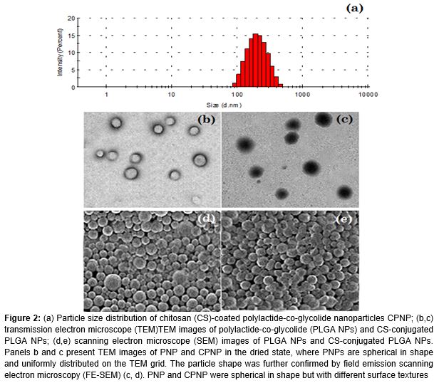
In the present study, a unique combination of DTX and ALN was used to treat osteosarcoma cells. For this purpose, CS was conjugated to the carboxylic group of PLGA and loaded with DTX and ALN. The dual-drug-loaded NPs were successfully prepared with high drug entrapment of 83.7 ± 1.23 and 71.89 % corresponding to 6.51 and 5.21 % drug loading of DTX and ALN. The particle size of the PLGA NPs (PNP) and CS-coated PLGA NPs (CPNP) was observed to be 165 ± 1.72 and 202 ± 4.2 nm respectively (a). The zeta potential of the NPs was evaluated to confirm the polymer substitution and stability. The zeta potential of the PNP was negatively charged at −23.67 ± 3.21 because of the carboxylic groups. The zeta potential changed to 21.37 ± 1.28 after coating with cationic polymer with an active amine functional group.
Morphology
The morphology of both NPs was evaluated using TEM and FE-SEM.
DSC thermograms
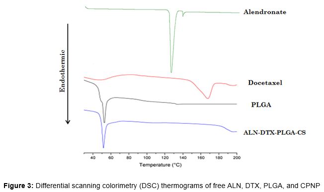
DSC was performed to assess the physical state of the drug after encapsulation in the NP as the physical state could influence the in vitro and in vivo properties of the drug. shows the DSC thermograms of pure DTX, ALN, PNP, and CPNP.
The melting endothermic peak of pure DTX appeared at 173 °C, while that of ALN appeared at 128 °C, indicating its sharp crystalline nature. The melting peak of both drugs was not detected in the NP formulation, indicating that the drugs were successfully incorporated into the NPs and present in the molecular dispersed and amorphous forms. It is worth noting that the crystalline form of the drug is not active and is not effective or stable in the systemic circulation. The amorphous form of the drug, however, circulates freely [13].
In vitro drug release
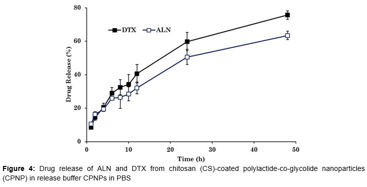
The release of DTX and ALN from CPNP was performed in PBS (pH 7.4) to simulate the physiological conditions of the human body. An initial release of approximately ~30 % from the NPs was observed in the first 8 h, suggesting that a significant portion of the drug was present on the surface of the NP, which was released upon contact with the medium (). Following this initial release, a more sustained release of the drug was observed for up to 48 h, demonstrating the capacity of the carrier to withhold the drug while in systemic circulation.
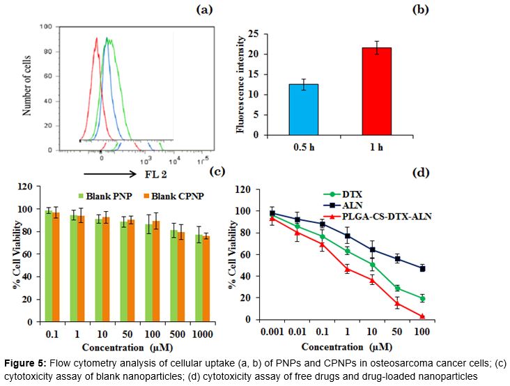
Cellular uptake
Flow cytometry analysis was performed to confirm the cellular uptake of CPNPs in MG-63 cancer cells. To accomplish this, the drug was replaced with the fluorescent probe Rhodamine B. A remarkable increase in fluorescence intensity was observed after 30-min incubation (a, b). Cellular uptake increased further when the incubation time was extended to 1 h.
In vitro cytotoxicity
The in vitro cytotoxicity of blank PLGA NPs and blank CS-coated PLGA NPs was investigated in MG-63 cancer cells. Cancer cells were exposed to the polymers (0.1 – 1,000 µg/mL). Blank NP treatment resulted in high cell viability, suggesting that the polymers have excellent biocompatibility (c, d). The lack of carrier cytotoxicity is advantageous for cancer cell targeting. MG-63 cells were also treated with free DTX, ALN, and DTX/ALN-loaded combined NPs. Both DTX and ALN treatment resulted in typical concentration-dependent cytotoxicity after 24 h incubation. Moreover, DTX was more effective than ALN in killing cancer cells.
Discussion
First, DTX- and ALN-loaded PLGA NPs were prepared, and the carboxylic groups of PLGA NPs were conjugated to CS by an EDC/NHS reaction. PLGA NPs have a reduced affinity for cancer cells due to the negatively charged carboxyl groups on the PLGA surface. CS was therefore introduced to modify the surface charge, and improve chemotherapeutic efficacy. The positively charged NP system thus increased cellular uptake efficiency. The increase in the size of NP was attributed to the presence of the high molecular weight CS on the NP surface. It also indicated a definite deposition of mass on the NPs. The zeta potential changed to a positive charge after coating with a cationic polymer with an active amine functional group. The presence of this positive charge was expected to enhance the interaction between the cancer cells and NPs and facilitate cellular uptake [12].
The spherical nature of the NP was attributed to the self-assembly of the polymer in the presence of surfactant. CPNPs are also spherical particles with monodispersion, although the overall particle size is larger. The greyish shell on the NP surface might have been the CS coating, which increased the particle size. PNPs initially consisted of a smooth surface, while the CS coating slightly roughened the surface. Nevertheless, a spherical-shaped particle in the nanodimension is ideal for cancer tissue targeting.
Importantly, no significant difference in the release pattern of DTX and ALN was observed, indicating that the substitution of CS on the NP surface did not influence drug release from the delivery system [13]. The release data were fit to mathematical models, and the release kinetics were confirmed by fitting to the Higuchi model (r = 0.999), with the release profile indicating a diffusion-based release. A controlled release system in which the drug is released to the tumour tissue at a steady rate is of significant importance.
High cellular uptake efficiency is also necessary for the successful delivery of the drug to the tumour. Our results suggest that positively charged nanocarriers could be efficiently internalized in cancer cells. DTX/ALN-loaded CPNPs exhibited a superior cancer killing effect compared with either drug alone. Interestingly, introduction of DTX/ALN-loaded CPNPs resulted in only 45 % cell viability, indicating its cytotoxicity. The IC50 value, or the concentration required to kill 50 % of cancer cells, was investigated: the IC50 value of DTX/ALN-loaded CPNPs was nearly 20 times lower than that of the free drugs [14].
Conclusion
A unique DTX- and ALN-conjugated CS-coated PLGA nanoparticle formulation was prepared and evaluated for its potential in targeting osteosarcoma cells. The two-drug-loaded NP showed a superior cancer killing effect in MG-63 cancer cells and was safe and biocompatible. Combined loading of DTX and ALN increased the therapeutic efficacy in osteosarcoma treatment. Thus, combinatorial drug regimens using nanocarriers hold promise for the effective treatment of osteosarcoma.
Declarations
Acknowledgement
References
Archives


News Updates